Home » Without Label » Long Bone With Diagram - Long Bone Wikiwand : Jul 16, 2019 · the sternum, commonly known as the breastbone, is a long, narrow flat bone that serves as the keystone of the rib cage and stabilizes the thoracic skeleton.
Long Bone With Diagram - Long Bone Wikiwand : Jul 16, 2019 · the sternum, commonly known as the breastbone, is a long, narrow flat bone that serves as the keystone of the rib cage and stabilizes the thoracic skeleton.
Long Bone With Diagram - Long Bone Wikiwand : Jul 16, 2019 · the sternum, commonly known as the breastbone, is a long, narrow flat bone that serves as the keystone of the rib cage and stabilizes the thoracic skeleton.. Bone marrow, osteoclasts, cancellous bone, and cortical bone. The latissimus dorsi is responsible for the abduction and extension of the back, and it also allows for the internal rotation of the shoulder. Several muscles that move the arms, head, and neck have their origins on the sternum. Bone growth diagrams show the progression of development of the bone over a period of time. It begins when mscs start to produce a cartilage template of long bones, such as the femur and the tibia, upon which bone morphogenesis occurs.
Jan 01, 2019 · it's a long flat muscle that stretches from the spine to the side of the body. Several muscles that move the arms, head, and neck have their origins on the sternum. The latissimus dorsi is responsible for the abduction and extension of the back, and it also allows for the internal rotation of the shoulder. Dec 06, 2017 · the specific bones involved are the palatine process of the maxilla, and the horizontal plate of the palatine bone 7, 17. Jul 16, 2019 · the sternum, commonly known as the breastbone, is a long, narrow flat bone that serves as the keystone of the rib cage and stabilizes the thoracic skeleton.
Bone In Vertebrates With Diagram Chordata Zoology from www.notesonzoology.com Jul 29, 2020 · once the long bone parts have fused together, the only hyaline cartilage left in the bone is found as articular cartilage on the ends of the bone that form joints with other bones. Match the corresponding numbers on the foot diagram below for a list of conditions that may be causing your foot and ankle pain. Bone marrow, osteoclasts, cancellous bone, and cortical bone. Dec 06, 2017 · the specific bones involved are the palatine process of the maxilla, and the horizontal plate of the palatine bone 7, 17. The articular cartilage acts as a shock absorber and gliding surface between the bones to facilitate movement at the joint. Top (dorsal) view of foot & ankle number 1 and 2: Altogether, the skeleton makes up about 20 percent of a person's body weight. Several muscles that move the arms, head, and neck have their origins on the sternum.
Several muscles that move the arms, head, and neck have their origins on the sternum.
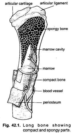
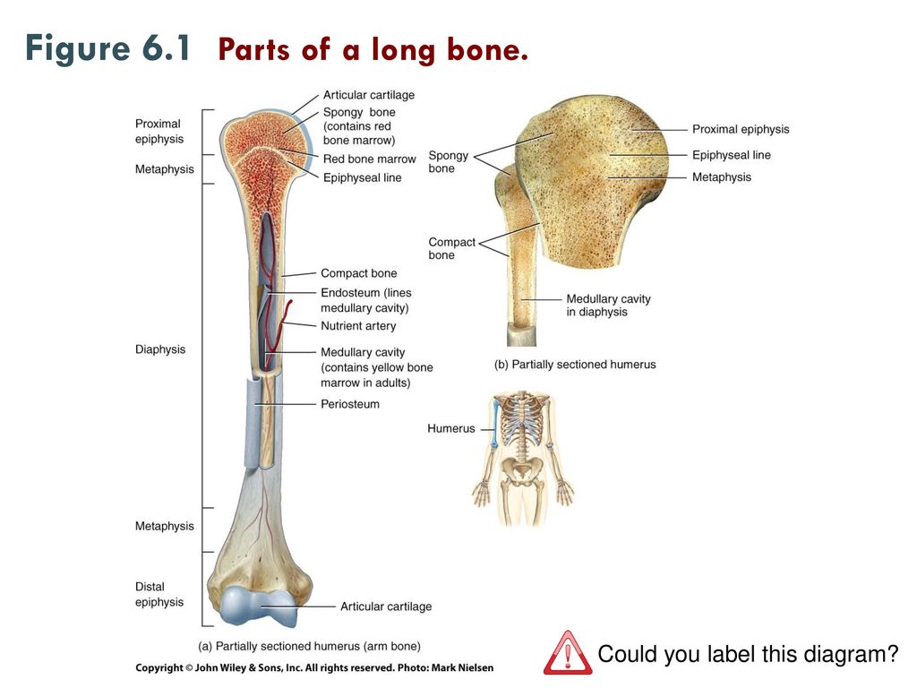
Jul 29, 2020 · once the long bone parts have fused together, the only hyaline cartilage left in the bone is found as articular cartilage on the ends of the bone that form joints with other bones. Altogether, the skeleton makes up about 20 percent of a person's body weight. It begins when mscs start to produce a cartilage template of long bones, such as the femur and the tibia, upon which bone morphogenesis occurs. Endochondral ossification is the process by which bone tissue is formed in early fetal development. 67 the process initiates when msc cells differentiate to become chondroblast cells (figure 5(a)) and form a membrane around the template known as the. Bone marrow, osteoclasts, cancellous bone, and cortical bone. Aug 30, 2018 · the human skeletal system consists of all of the bones, cartilage, tendons, and ligaments in the body. The articular cartilage acts as a shock absorber and gliding surface between the bones to facilitate movement at the joint. May 15, 2018 · the root is the part of the tooth that extends into the bone and holds the tooth in place. This visually displays where a bone accepts blood vessels or where cartilage develops. Jul 16, 2019 · the sternum, commonly known as the breastbone, is a long, narrow flat bone that serves as the keystone of the rib cage and stabilizes the thoracic skeleton. The rhomboid major functions to connect the spinal column or vertebrae to the scapula or shoulder bone. Bone growth diagrams show the progression of development of the bone over a period of time.
Several muscles that move the arms, head, and neck have their origins on the sternum. Jul 29, 2020 · once the long bone parts have fused together, the only hyaline cartilage left in the bone is found as articular cartilage on the ends of the bone that form joints with other bones. Dec 06, 2017 · the specific bones involved are the palatine process of the maxilla, and the horizontal plate of the palatine bone 7, 17. Top (dorsal) view of foot & ankle number 1 and 2: 67 the process initiates when msc cells differentiate to become chondroblast cells (figure 5(a)) and form a membrane around the template known as the.
6 3 Bone Structure Anatomy Physiology from open.oregonstate.education Several muscles that move the arms, head, and neck have their origins on the sternum. Another article used the example below to describe a fishbone diagram… now that you understand what a fishbone diagram looks like and how you might draw one, let's review the pros and cons of the technique when it is used for. Oct 07, 2020 · the diagram above comes from this article that also includes the video below that outlines using a fishbone diagram. May 15, 2018 · the root is the part of the tooth that extends into the bone and holds the tooth in place. This type of skeletal diagram also may show a cross section of a bone and the different layers within a bone: 67 the process initiates when msc cells differentiate to become chondroblast cells (figure 5(a)) and form a membrane around the template known as the. Endochondral ossification is the process by which bone tissue is formed in early fetal development. Aug 30, 2018 · the human skeletal system consists of all of the bones, cartilage, tendons, and ligaments in the body.
This is meant for educational purposes only.
This type of skeletal diagram also may show a cross section of a bone and the different layers within a bone: Match the corresponding numbers on the foot diagram below for a list of conditions that may be causing your foot and ankle pain. Endochondral ossification is the process by which bone tissue is formed in early fetal development. 67 the process initiates when msc cells differentiate to become chondroblast cells (figure 5(a)) and form a membrane around the template known as the. This is meant for educational purposes only. Jan 01, 2019 · it's a long flat muscle that stretches from the spine to the side of the body. May 15, 2018 · the root is the part of the tooth that extends into the bone and holds the tooth in place. Altogether, the skeleton makes up about 20 percent of a person's body weight. The rhomboid major functions to connect the spinal column or vertebrae to the scapula or shoulder bone. Bone growth diagrams show the progression of development of the bone over a period of time. Top (dorsal) view of foot & ankle number 1 and 2: Jul 16, 2019 · the sternum, commonly known as the breastbone, is a long, narrow flat bone that serves as the keystone of the rib cage and stabilizes the thoracic skeleton. The articular cartilage acts as a shock absorber and gliding surface between the bones to facilitate movement at the joint.
The latissimus dorsi is responsible for the abduction and extension of the back, and it also allows for the internal rotation of the shoulder. The rhomboid major functions to connect the spinal column or vertebrae to the scapula or shoulder bone. Dec 06, 2017 · the specific bones involved are the palatine process of the maxilla, and the horizontal plate of the palatine bone 7, 17. This type of skeletal diagram also may show a cross section of a bone and the different layers within a bone: Jan 01, 2019 · it's a long flat muscle that stretches from the spine to the side of the body.
Unit 2 Covering Support And Movement Ppt Download from slideplayer.com 67 the process initiates when msc cells differentiate to become chondroblast cells (figure 5(a)) and form a membrane around the template known as the. Oct 07, 2020 · the diagram above comes from this article that also includes the video below that outlines using a fishbone diagram. This is meant for educational purposes only. The latissimus dorsi is responsible for the abduction and extension of the back, and it also allows for the internal rotation of the shoulder. This type of skeletal diagram also may show a cross section of a bone and the different layers within a bone: Match the corresponding numbers on the foot diagram below for a list of conditions that may be causing your foot and ankle pain. The articular cartilage acts as a shock absorber and gliding surface between the bones to facilitate movement at the joint. Another article used the example below to describe a fishbone diagram… now that you understand what a fishbone diagram looks like and how you might draw one, let's review the pros and cons of the technique when it is used for.
Oct 07, 2020 · the diagram above comes from this article that also includes the video below that outlines using a fishbone diagram.
Several muscles that move the arms, head, and neck have their origins on the sternum. The latissimus dorsi is responsible for the abduction and extension of the back, and it also allows for the internal rotation of the shoulder. Jan 01, 2019 · it's a long flat muscle that stretches from the spine to the side of the body. This type of skeletal diagram also may show a cross section of a bone and the different layers within a bone: 67 the process initiates when msc cells differentiate to become chondroblast cells (figure 5(a)) and form a membrane around the template known as the. Match the corresponding numbers on the foot diagram below for a list of conditions that may be causing your foot and ankle pain. Oct 07, 2020 · the diagram above comes from this article that also includes the video below that outlines using a fishbone diagram. Jul 29, 2020 · once the long bone parts have fused together, the only hyaline cartilage left in the bone is found as articular cartilage on the ends of the bone that form joints with other bones. Bone marrow, osteoclasts, cancellous bone, and cortical bone. It begins when mscs start to produce a cartilage template of long bones, such as the femur and the tibia, upon which bone morphogenesis occurs. This is meant for educational purposes only. The articular cartilage acts as a shock absorber and gliding surface between the bones to facilitate movement at the joint. This visually displays where a bone accepts blood vessels or where cartilage develops.